Our Products
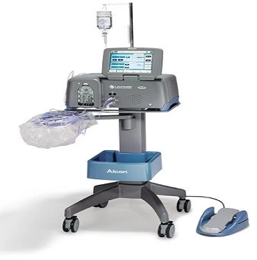
Phaco Machine
Phacoemulsification is a modern cataract surgery in which the eye's internal lens is emulsified with an ultrasonic hand piece and aspirated from the eye. Phaco- emulsification is the technique by which cataract is broken into small pieces and removed from the eye using ultrasound with the help of a special phaco equipment. All phaco machines consist of a computer to generate electrical signals and a transducer to turn these electronic signals into mechanical energy. The energy thus produced is passed through a hollow needle and is controlled within the eye to overcome the inertia of the lens and emulsify it. During surgery, the phaco machine keeps track of the average phaco power, given as a percentage of maximum, as well as the total time during which phaco ultrasonic power was delivered. The phaco machine broadly consists of :
- Console
- Foot pedal
- Hand piece and their connections
From the time of you procure equipment, precautions and systems should be put in place to ensure long service life and competency. And, if you are seeking professional assistance for doing so, then YE is the right place to contact. With our phaco machine repairing services, we work in coordination with clients and take preventive measures to keep phaco machine serviceable. For minor operational glitches, we provide on-site solutions and for major hiccups, we bring the machine to our workshop and repair it skillfully. We are based in Neemuch (Madhya Pradesh, India); clients can contact us anytime for further discussion.
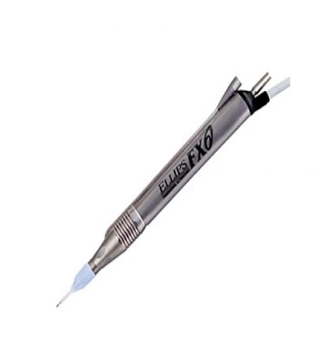
Phaco Hand-Piece
The Phaco Hand Piece contains the piezoelectric crystal, which is in contact with the tip. The tip is covered by a silicon sleeve. The infusion fluid flows between the tip and the sleeve cooling the former. Power of the machine depends upon the stroke length and the frequency remains fixed. There are two openings on the sleeve for the exit of this fluid, which should be kept perpendicular to the tip bevel. The proximal end of hand piece is connected to the console with an electric cord. There are two more connections: one each for the irrigation tubing and for connecting the aspiration system. Functioning of the probe is to deliver the energy which cuts the hard part of cataract which is the nucleus. The mechanism of working is by:
- Jackhammer effect
- Cavitation impact
- Acoustic wave of fluid
With 5+ years of experience and industry knowledge, YE is providing all sorts of solutions related to phaco handpiece repairing needs. We are based in Neemuch (Madhya Pradesh, India) and serving throughout the region. Ours is a team of professionals and technicians, who work as per clients schedule and provide precise solutions in less time. Besides, we keep a stock of phaco handpiece suitable for all makes and models. Our service charges are reasonable and depends on the job work.
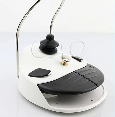
Phacto Foot Pedal
Foot pedal control is the most important aspect of phaco. Though the foot pedal of each machine may have a different design, it essentially consists of main central part and sidekicks. The main part of the foot pedal controls infusion, aspiration and phaco power. The entire distance that the foot pedal traverses is divided by 2 dentations into 3 excursions-
- I (irrigation only),
- IA (infusion and aspiration)
- IAP (infusion, aspiration and phaco)
Get in touch with us if you are seeking professional assistance that can undertake the repairing work of phaco machine foot pedal. YE is a professionally managed service provider and actively involved in providing phaco machine foot pedal repairing services to the clients in the surrounding regions of Neemuch (Madhya Pradesh, India). Our services are rendered by a group of technicians and as per the stated industry norms. With us, you are free to schedule repair as per your convenience; we assure to work accordingly.
Vitrectomy Cutter
Vitrectomy is a type of eye operation. A doctor removes the vitreous, a jelly-like fluid inside your eye, and replaces it with a saline solution. For this operation, Vitrectomy Cutter/ Vitreous Cutter is used. It allows high-speed cutting of formed vitreous and controlled removal of the vitreous gel with low suction. It uses electric energy to drive the motor, heavier. It Utilizes pressurized air pulses to drive the diaphragm and inner tube forward, light weight. The vitreous cutter is connected to the vitrectomy machine through two lines. First goes from the cutter to the cutting drive on the machine. The second line is the aspiration line from the vitreous cutter to the vitrectomy machine. Higher the cut rate, the smaller the amount of vitreous (“bite size”) aspirated into the cutter, reducing both vitreous and retinal traction.
- Guillotine-type mechanism, which in most cases is driven pneumatically by the vitrectomy machine.
- Rotatory cutters the inner tube rotates within the outer tube to cut the vitreous.
- Oscillatory cutters are similar to rotatory type, but the rotation is not 360°. They rotate 180° to one side to cut the vitreous and again 180° to other side again
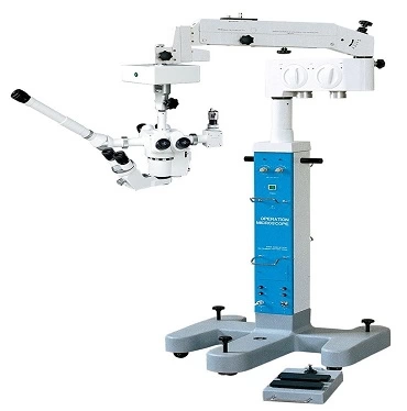
Microscope
A microscope is a scientific instrument. It makes small objects look larger. This lets people see the small things. People who use microscopes frequently in their jobs include doctors and scientists. Slit lamp microscopes are used by ophthalmologists to directly examine the eyes of a patient under the magnification of a binocular microscope by creating a stereoscopic, erect image. The slit lamp CS-1 microscope produces a narrow beam of intense light that can illuminate the patient's cornea, aqueous humour, crystalline lens, anterior vitreous layer, or other transparent ocular tissue. A form of oblique microscopy, the slit lamp microscope uses an imaging technique similar to optical sectioning. Interchangeable paired 10x and 15x eyepieces fit the binocular tubes of the CS-1 bio microscope. Thus, the ophthalmologist controls the magnification during an eye examination.
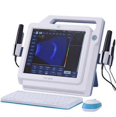
Ultra Sonography
Ultrasonography (USG) is application of medical with ultrasound-based imaging diagnostic technique used to visualize internal organs, their size, structure and their pathological lesions. Ultrasonography is widely utilized in medicine, primarily in gastroenterology, cardiology, gynaecology and obstetrics, urology and endocrinology Ultrasonography is generally considered a "safe" imaging modality. However slight detrimental effects have been occasionally observed (see below). Ultrasound information can be displayed in several ways
- A-mode: This display mode is the simplest; signals are recorded as spikes on a graph. This type of ultrasonography is used for ophthalmologic scanning.
- B-mode (gray-scale): It is commonly used to evaluate the developing foetus and to evaluate organs, including the liver, spleen, kidneys, thyroid gland, testes, breasts, and prostate gland. It is fast enough to show real-time motion, such as the motion of the beating heart or pulsating blood vessels. Real- time imaging provides anatomic and functional information.
- M-mode: This mode is used to image moving structures; signals reflected by the moving structures are converted into waves that are displayed continuously across a vertical axis.
Taking care of your equipment is the best way to ensure its longevity and competency. If you are seeking professional assistance that can handle this affair assiduously, then get in touch with us. Ours is a Neemuch (Madhya Pradesh, India) based service provider company and providing Ultra Sonography machine repairing services to the clients in the surrounding regions. Executed by technicians, our services are available for all makes and models. Our service charges are reasonable and we provide solutions as per clients’ schedule.
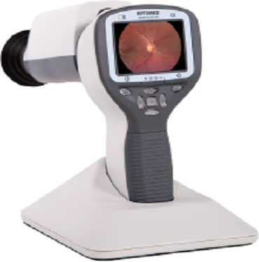
Fundus Camera
A fundus camera or retinal camera is a specialized low power microscope with an attached camera designed to photograph the interior surface of the eye, including the retina, retinal vasculature, optic disc, macula, and posterior pole (i.e. the fundus).The main structures that can be visualized on a fundus photo are the central and peripheral retina, optic disc and macula. Fundus photography can be performed with coloured filters, or with specialized dyes including fluorescein and indo cyanine green.
Yag and Green Ledger
A Combination YAG/Green Laser is generally considered a multipurpose laser for use in treating cataracts and glaucoma. What makes this product unique is that it allows the surgeon to switch between different types of lasers that are all housed in one piece of equipment, offering space, energy and time savings. The combination laser systems are normally built over a slit lamp, and practitioners can move between the lasers by the push of a button.
- Designed to reduce laser lens reflections while maintaining laser-beam integrity.
- The only combo system with two individual systems that act like one — without compromise.
- Facile connectivity between the YC-1800 and GYC-1000 for a wider range of treatment options.
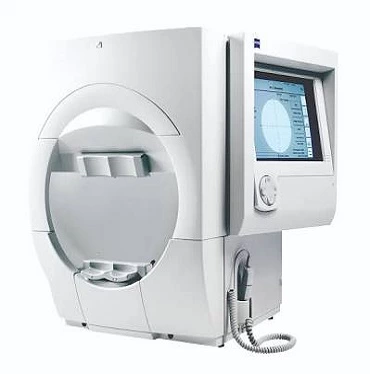
Field Analyser
Humphrey field analyser (HFA), is a tool for measuring the human visual field. It is used by optometrists, orthoptists and ophthalmologists, particularly for detecting monocular visual field. The results of the Analyser identify the type of vision defect. Therefore, it provides information regarding the location of any disease processes or lesion(s) throughout the visual pathway. This guides and contributes to the diagnosis of the condition affecting the patient's vision. These results are stored and used for monitoring the progression of vision loss and the patient's condition.
What people say about us

Tanishk Agrawal
"Their Service is very quick and must say I liked their work style"
Yogesh Rawat
"Quality is the only key for their growth."
Palash Khandelwal
"It is experienced, cheaper and provide quality. I seriously love their work."
Adil Hashmi
"Compared to other,It has more professional and quick."
Archit Guhey
"Understand the problem of Customers and find it's solution as fast as possbile"
Ojasvee Kirar
"Very Good Customer Care Service."
Ibrahim Arif
"Using Quality material while repairing makes them different"